Upper Leg Tendon Anatomy - Muscles Of The Leg Part 2 Anterior And Lateral Compartments Anatomy Tutorial Youtube : This may result in tendon subluxation;
Upper Leg Tendon Anatomy - Muscles Of The Leg Part 2 Anterior And Lateral Compartments Anatomy Tutorial Youtube : This may result in tendon subluxation;. When a muscle contracts, the tendon pulls on the bone causing the joint to move. Relation of the tendon of flexor hallucis longus to the. Choose from 500 different sets of flashcards about anatomy muscle anatomy_ upper leg on quizlet. Related online courses on physioplus. The triceps tendon is wider than most of the other tendons in the upper extremity.
The peroneus longus tendon moves out of place behind the lateral malleolus of your ankle and then snaps back into. The pads of the machine are situated at the achilles tendon. The achilles tendon or heel cord, also known as the calcaneal tendon, is a tendon at the back of the lower leg, and is the thickest in the human body. Related online courses on physioplus. 3d illustration back fit strong human anatomy.
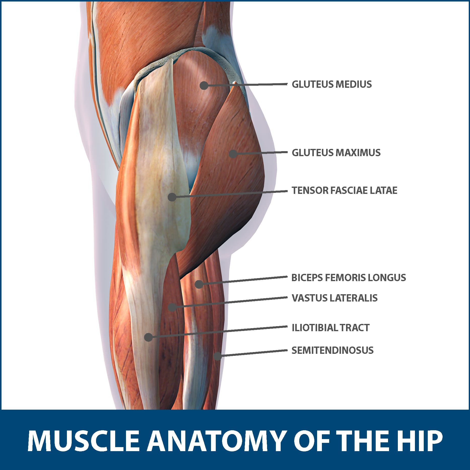
Hip Muscle Strains Info Florida Orthopaedic Institute from www.floridaortho.com We speak of the upper extremities (arms) and the lower extremities (legs). Muscle/tendon inflammation and pain along anterio… Hands are outstretched, holding onto the handles of the bench. They are innervated by the tibial nerve, a terminal branch of the sciatic nerve. Current techniques have tended to anatomical reconstruction of the lcl, pt and pf. What are the functions of patella. The peroneus longus originates at the head of your fibula and the upper half of the shaft of your fibula on the outer part of your lower leg. Related posts of muscle anatomy upper leg.
They have blood vessels and cells to maintain tendon health and repair injured tendon.
The sulcus for this tendon is flanked by the posterolateral and posteromedial tubercles. Its muscle belly is on the back aspect of the upper arm. What are the functions of patella. This may result in tendon subluxation; The tendons for these muscles begin at your ischial tuberosity, or ischium (the. Mnemonics that can be used to remember the anatomy of the ankle tendons from anterior to posterior as they pass posteriorly to the medial malleolus of the tibia under the flexor retinaculum in the tarsal tunnel include: The posterior talofibular ligament is attached to the posterolateral tubercle, which is larger and more prominent than the posteromedial tubercle. 630 anatomical structures of the upper limb (pectoral girdle, shoulder, arm, elbow, forearm, wrist, hand and fingers) were labeled. This mri wrist coronal cross sectional anatomy tool is absolutely free to use. Lateral (fibular) collateral ligament (fcl) upper part middle part lower part popliteus tendon (pt) upper part i. .16 penile numbness and perineum tenderness.18 any suggested exercises or stretches?.22 leg musculature 209 elbow tendonitis and saddle sores. Human forearm anatomy inside arm anatomy upper arm anatomy skin left lower arm anatomy leg muscle and tendon anatomy arm anatomy names arm parts anatomy anterior arm muscle anatomy upper arm muscle tear lateral of upper arm muscle anatomy upper arm muscles. The triceps tendon is wider than most of the other tendons in the upper extremity.
The achilles tendon or heel cord, also known as the calcaneal tendon, is a tendon at the back of the lower leg, and is the thickest in the human body. Lie prone on a hamstring curl machine. Hands are outstretched, holding onto the handles of the bench. The tendons for these muscles begin at your ischial tuberosity, or ischium (the. Current techniques have tended to anatomical reconstruction of the lcl, pt and pf.
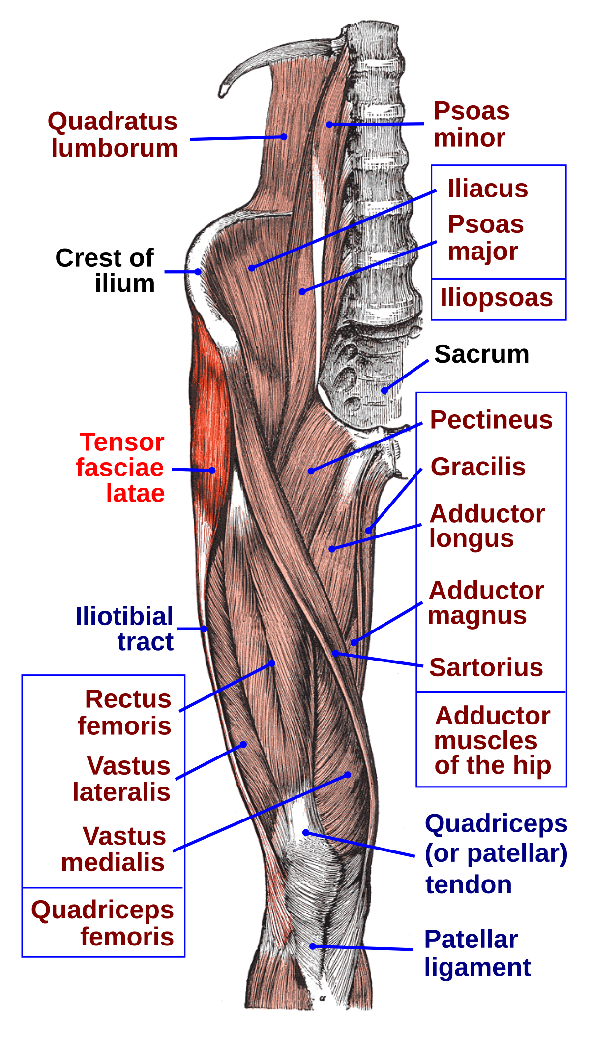
Tensor Fasciae Latae Muscle Wikipedia from upload.wikimedia.org Lateral (fibular) collateral ligament (fcl) upper part middle part lower part popliteus tendon (pt) upper part i. Collectively, the muscles in this area plantarflex and invert the foot. We study anatomy at the practical anatomy class we study the human body. Superficial veins of upper limb , anatomy : Current techniques have tended to anatomical reconstruction of the lcl, pt and pf. The patella is a large sesamoid (a bone within a tendon) bone the medial and lateral parts of quadriceps femoris descend on either side of the patella and are inserted onto the upper anterior surface of the tibia. This may result in tendon subluxation; After completion of this video, you will be able to identify and discuss some features of the calf and sole of the foot:
The tendons for these muscles begin at your ischial tuberosity, or ischium (the.
Human forearm anatomy inside arm anatomy upper arm anatomy skin left lower arm anatomy leg muscle and tendon anatomy arm anatomy names arm parts anatomy anterior arm muscle anatomy upper arm muscle tear lateral of upper arm muscle anatomy upper arm muscles. Anatomy of leg and foot human muscular system stock vector.,category:anatomy of the human leg,muscles of the leg and foot classic human anatomy in motion: In this upper leg tutorial, i go over all the major points of the upper leg to take your sculpting skills. Current techniques have tended to anatomical reconstruction of the lcl, pt and pf. Its muscle belly is on the back aspect of the upper arm. Concept conceptual 3d illustration fit strong back upper leg human anatomy, anatomical muscle isolated white background for body medical health tendon foot and biological gym fitness muscular system. The achilles tendon or heel cord, also known as the calcaneal tendon, is a tendon at the back of the lower leg, and is the thickest in the human body. The pads of the machine are situated at the achilles tendon. It serves to attach the plantaris, gastrocnemius (calf) and soleus muscles to the calcaneus (heel) bone. Lateral (fibular) collateral ligament (fcl) upper part middle part lower part popliteus tendon (pt) upper part i. Related posts of muscle anatomy upper leg. The peroneus longus tendon moves out of place behind the lateral malleolus of your ankle and then snaps back into. Collectively, the muscles in this area plantarflex and invert the foot.
They have blood vessels and cells to maintain tendon health and repair injured tendon. Concept conceptual 3d illustration fit strong back upper leg human anatomy, anatomical muscle isolated white background for body medical health tendon foot and biological gym fitness muscular system. There is no real division between the core and the upper leg; The achilles tendon or heel cord, also known as the calcaneal tendon, is a tendon at the back of the lower leg, and is the thickest in the human body. Tendons are thick bands of tissue that connect muscles to bone.
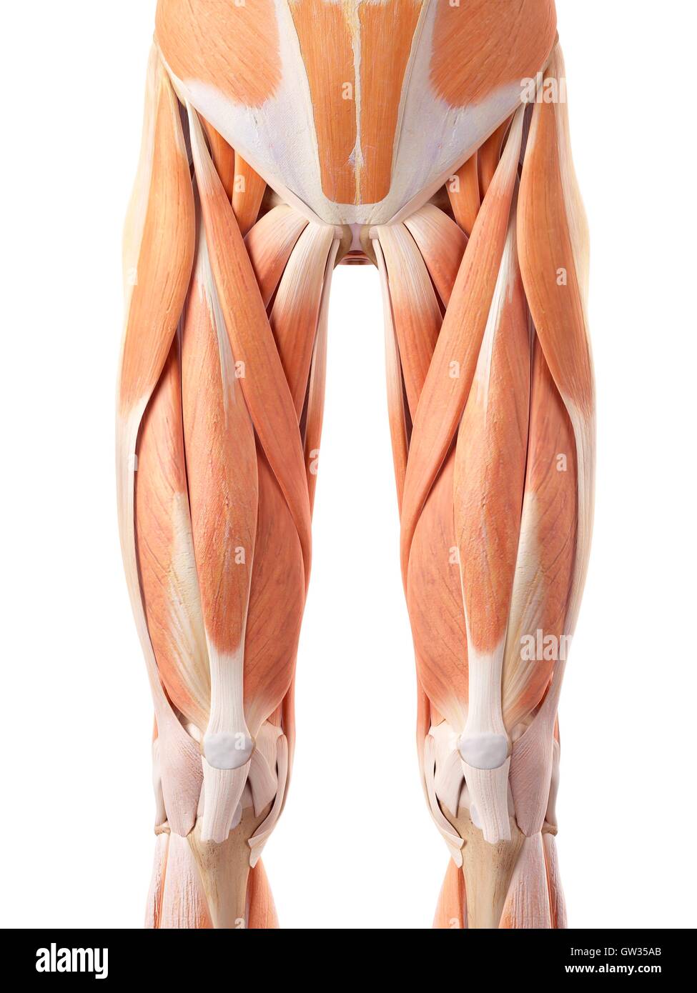
Page 3 Upper Leg Muscles High Resolution Stock Photography And Images Alamy from c8.alamy.com Current techniques have tended to anatomical reconstruction of the lcl, pt and pf. ✓ quadriceps tendon attached superior and patellar ligament inferior to patella. The achilles tendon or heel cord, also known as the calcaneal tendon, is a tendon at the back of the lower leg, and is the thickest in the human body. The triceps tendon is wider than most of the other tendons in the upper extremity. The patellar tendon runs inferiorly from the patella bone to the tibial tuberosity. Palmar region , arteries (illustrations: Tendons are cords made of tough tissue, and they work as special connector pieces between bone and muscle. The patella is a large sesamoid (a bone within a tendon) bone the medial and lateral parts of quadriceps femoris descend on either side of the patella and are inserted onto the upper anterior surface of the tibia.
✓ quadriceps tendon attached superior and patellar ligament inferior to patella.
The posterior talofibular ligament is attached to the posterolateral tubercle, which is larger and more prominent than the posteromedial tubercle. Concept 3d illustration back upper leg human anatomy. An anatomical and biomechanical study. Arrangement of tendons of tibialis posterior, flexor digitorum longus, flexor hallucis longus; This mri wrist coronal cross sectional anatomy tool is absolutely free to use. .16 penile numbness and perineum tenderness.18 any suggested exercises or stretches?.22 leg musculature 209 elbow tendonitis and saddle sores. Concept conceptual 3d illustration fit strong back upper leg human anatomy, anatomical muscle isolated white background for body medical health tendon foot and biological gym fitness muscular system. Muscle/tendon inflammation and pain along anterio… Tendons are cords made of tough tissue, and they work as special connector pieces between bone and muscle. ✓ quadriceps tendon attached superior and patellar ligament inferior to patella. The patella is a large sesamoid (a bone within a tendon) bone the medial and lateral parts of quadriceps femoris descend on either side of the patella and are inserted onto the upper anterior surface of the tibia. Mnemonics that can be used to remember the anatomy of the ankle tendons from anterior to posterior as they pass posteriorly to the medial malleolus of the tibia under the flexor retinaculum in the tarsal tunnel include: 630 anatomical structures of the upper limb (pectoral girdle, shoulder, arm, elbow, forearm, wrist, hand and fingers) were labeled.


0 Komentar